
File:Diseases of Swine 17-7.png

本预览的尺寸:724 × 599像素。 其他分辨率:290 × 240像素 | 580 × 480像素 | 928 × 768像素 | 1,237 × 1,024像素 | 1,550 × 1,283像素。
原始文件 (1,550 × 1,283像素,文件大小:2.12 MB,MIME类型:image/png)
| 描述 |
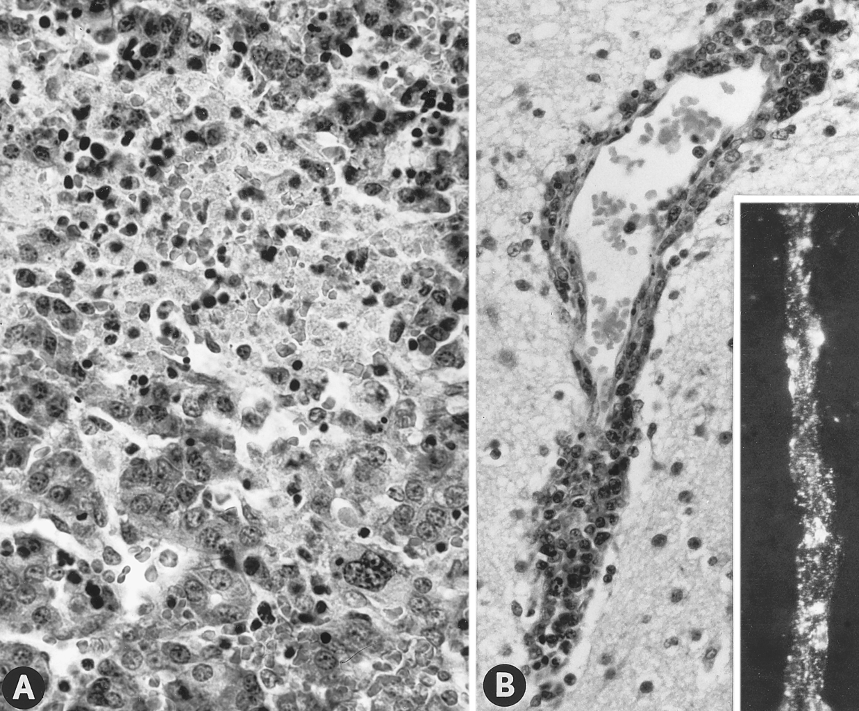
English: 17.7. Tissues of PPV-infected fetuses of gilts experimentally infected oronasally. (A) Necrotic focus in liver of live fetus of a gilt infected on day 40 of gestation and killed 42 days later; fetus had numerous macroscopic lesions (H&E; ×400). (B) Perivascular cuffing with mononuclear cells in cerebrum of live fetus, littermate of A; fetus had no macroscopic lesions (H&E; ×320). (Insert) Viral antigen associated with endothelium of cerebral vessel of fetus of a gilt infected on day 46 of gestation and killed 25 days later (IF microscopy; ×312.5). All fetuses were probably infected by intrauterine spread of PPV from transplacentally infected littermates. (Photographs A and B courtesy of T. T. Brown, Jr., National Animal Disease Center.) |
||
| 来源 | Diseases of Swine (8th edition) | ||
| 作者 | W. L. Mengeling | ||
| 授权 (二次使用本文件) |
|
According to its copyright statement, "Copyright is not claimed for Chapters 17, 23, 25, 31, and 64, which are in the public domain.".
文件历史
点击某个日期/时间查看对应时刻的文件。
| 日期/时间 | 缩略图 | 大小 | 用户 | 备注 | |
|---|---|---|---|---|---|
| 当前 | 2009年3月19日 (四) 11:07 | | 1,550 × 1,283(2.12 MB) | Dingar | {{Information |Description={{en|1=17.7. Tissues of PPV-infected fetuses of gilts experimentally infected oronasally. (A) Necrotic focus in liver of live fetus of a gilt infected on day 40 of gestation and killed 42 days later; fetus had numerous macroscop |
文件用途
没有页面使用本文件。
全域文件用途
以下其他wiki使用此文件:
- ca.wikipedia.org上的用途
- en.wikipedia.org上的用途
- en.wikisource.org上的用途
- zh.wikipedia.org上的用途